Diagram Of Shoulder / Standard Anatomic Total Shoulder Replacement Dr Gordon Groh. Inside the shoulder there are three joints; The acromioclavicular joint is formed by an articulation between the lateral end of the clavicle and the acromion process of the scapula. These cases may resolve prior to identifying a clear diagnosis or a specific diagnosis may become clear with time. It is one of the most mobile joints in the human body, at the cost of joint stability. Muscles of the shoulder :
The rotator cuff is a group of four muscles and tendons that surround the glenohumeral joint. Four of them are found on the anterior aspect of the shoulder, whereas the rest are located on the shoulder's posterior aspect and in the back. It is an extremely mobile joint, in which stability has been sacrificed for mobility. The labrum also serves as the attachment of a major tendon in the shoulder, the biceps tendon. These muscles form the outer shape of the shoulder and underarm.
Illustration Of The Bony Anatomy Of The Shoulder Joint Complex Download Scientific Diagram from www.researchgate.net This is the smallest rotator cuff muscle. Atlas of the anatomy of the joint of the shoulder on a ct arthrogram in axial, coronal, and sagittal sections, on a 3d images and on conventional. These muscles form the outer shape of the shoulder and underarm. The shoulder muscles and shoulder tendons involved with shoulder mobility include the four rotator cuff muscle and tendon pairs: Diagram of the shoulder, including the location of the rotator cuff. While seated or standing, lift the sore arm forward and to the side about thirty to 45 degrees. A diagram of an anatomic shoulder replacement—the plastic socket replaces the cup of the scapula (shoulder blade). The shoulder is a complex combination of bones and joints where many muscles act to provide the widest range of motion of any part of the body.
To further reinforce the shoulder, the four muscles of the rotator cuff extend from the scapula and surround the head of the humerus to both rotate the arm and prevent dislocation.
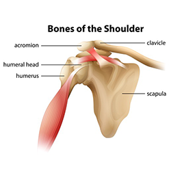
The supraspinatus is located on the upper part of the shoulder joint and is involved in abduction (arm raising). Beyond this, there is also a shoulder joint arrayed in a ball and socket formation, a rotator cuff, and various muscles like the deltoid muscle and the teres major muscle. The shoulder blade is called the scapula and the collarbone is called the clavicle. The shoulder is made up of two joints, the acromioclavicular joint and the glenohumeral joint. The shoulder is not a single joint, but a complex arrangement of bones, ligaments, muscles, and tendons that is better called the shoulder girdle. A metal ball component replaces the worn humeral head. This is why you need to get medical attention if you have shoulder pain—and the treatment is tailored to the cause, your overall health, and your level of activity. The socket of the shoulder joint is shallow, and the labrum gives the socket more depth, and thus more stability. Common rotator cuff injuries include rotator cuff tendonitis and rotator cuff strain, which is a partial or complete tear of the rotator cuff. What are common rotator cuff injuries? The list of muscles and their functions are presented below. This is the smallest rotator cuff muscle. Subscapularis, supraspinatus, infraspinatus and teres minor.
Shoulder pain has many different causes and treatments. Is the wear and tear of shoulder cartilage until bare bone is exposed. On the left is a standard (anatomic) shoulder arthroplasty. Smartdraw includes 1000s of professional healthcare and anatomy chart templates that you can modify and make your own. Subscapularis, supraspinatus, infraspinatus and teres minor.
Conventional Vs Reverse Total Shoulder Replacement Direct Orthopedic Care from www.directorthocare.com The components of the ball and cup are reversed on the right—a reverse shoulder replacement. Numerous muscles help stabilize the three joints of. The glenohumeral joint is where the ball (humeral head) and the socket (the glenoid) meet. These are located in the shoulder blade area, and each related tendon also attaches to the humerus. Muscles of the shoulder : Subscapularis, supraspinatus, infraspinatus and teres minor. Find symptoms,causes and treatments of joint disorders.for your health. To further reinforce the shoulder, the four muscles of the rotator cuff extend from the scapula and surround the head of the humerus to both rotate the arm and prevent dislocation.
These muscles form the outer shape of the shoulder and underarm.
Neck muscle anatomy mri 12 photos of the neck muscle anatomy mri neck muscle anatomy images, neck muscle anatomy pictures, neck muscle anatomy posterior, neck muscle anatomy ultrasound, neck muscles anatomy radiology, human muscles, neck muscle anatomy images, neck muscle anatomy pictures, neck muscle anatomy. Diagram of the shoulder, including the location of the rotator cuff. The acromioclavicular joint is where the acromion, part of the shoulder blade (scapula) and the collar bone (clavicle) meet. Ebraheim's educational animated video describes muscle anatomy of the shoulder girdle and anatomy of the shoulder joint.anatomy of the shoulder muscles a. The acromioclavicular joint is formed by an articulation between the lateral end of the clavicle and the acromion process of the scapula. While seated or standing, lift the sore arm forward and to the side about thirty to 45 degrees. Pronate your wrist so the palm of your hand faces down to the floor (as if you were trying to empty a glass of water). The shoulder joint is formed where the humerus (upper arm bone) fits into the scapula (shoulder blade), like a ball and socket. The supraspinatus is located on the upper part of the shoulder joint and is involved in abduction (arm raising). To further reinforce the shoulder, the four muscles of the rotator cuff extend from the scapula and surround the head of the humerus to both rotate the arm and prevent dislocation. The shoulder girdle includes three bones—the scapula, clavicle and humerus. The rotator cuff is a group of four muscles and tendons that surround the glenohumeral joint. Beyond this, there is also a shoulder joint arrayed in a ball and socket formation, a rotator cuff, and various muscles like the deltoid muscle and the teres major muscle.
These cases may resolve prior to identifying a clear diagnosis or a specific diagnosis may become clear with time. The muscles of the shoulder support and produce the movements of the shoulder girdle.they attach the appendicular skeleton of the upper limb to the axial skeleton of the trunk. These are located in the shoulder blade area, and each related tendon also attaches to the humerus. The shoulder has about eight muscles that attach to the scapula, humerus, and clavicle. Smartdraw includes 1000s of professional healthcare and anatomy chart templates that you can modify and make your own.
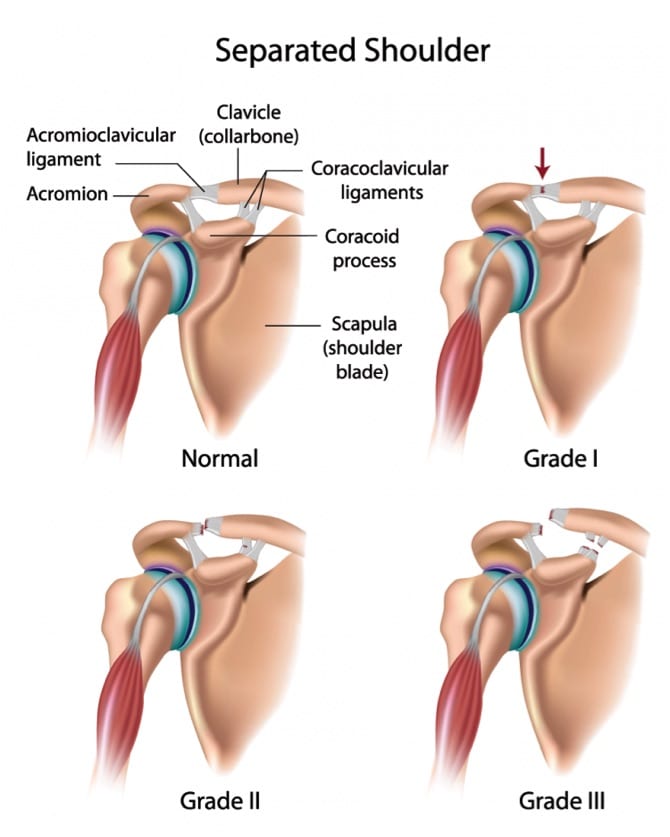
3 Grades Of Separated Shoulder Diagnosis And Treatment from charmaustin.com Just like the keystone in an archway. A diagram of an anatomic shoulder replacement—the plastic socket replaces the cup of the scapula (shoulder blade). It isn't easy to know the difference between different types of shoulder pain, like a frozen shoulder, shoulder blade pain, or symptoms of a rotator cuff tear. Shoulder pain is a common symptom in primary care. The shoulder is made up of two joints, the acromioclavicular joint and the glenohumeral joint. Sechrest, md narrates an animated tutorial on the basic anatomy of the shoulder. The top of the humerus is shaped like a ball. Find symptoms,causes and treatments of joint disorders.for your health.
It is an extremely mobile joint, in which stability has been sacrificed for mobility.
The rotator cuff is a group of four muscles and tendons that surround the glenohumeral joint. Diagram of shoulder pain : While seated or standing, lift the sore arm forward and to the side about thirty to 45 degrees. It is a flat, gliding joint. This is the main muscle that lets you rotate and extend your shoulder. These cases may resolve prior to identifying a clear diagnosis or a specific diagnosis may become clear with time. Numerous muscles help stabilize the three joints of. The bones of the pectoral girdle (clavicle and scapula) provide increased mobility to the. The anatomy of the shoulder. It is one of the most mobile joints in the human body, at the cost of joint stability. Four of them are found on the anterior aspect of the shoulder, whereas the rest are located on the shoulder's posterior aspect and in the back. Smartdraw includes 1000s of professional healthcare and anatomy chart templates that you can modify and make your own. Sechrest, md narrates an animated tutorial on the basic anatomy of the shoulder.
Share :
Post a Comment
for "Diagram Of Shoulder / Standard Anatomic Total Shoulder Replacement Dr Gordon Groh"


Post a Comment for "Diagram Of Shoulder / Standard Anatomic Total Shoulder Replacement Dr Gordon Groh"